Content
Cattle mycoplasmosis is a difficult to diagnose and, most importantly, an intractable disease that causes significant economic damage to farmers. The causative agent is widespread throughout the world, but due to the successful "masking" the disease is often misidentified.
What is this disease "mycoplasmosis"
The causative agent of the disease is a unicellular organism that occupies an intermediate position between bacteria and viruses. Representatives of the genus Mycoplasma are capable of independent reproduction, but they do not have the cell membrane inherent in bacteria. Instead of the latter, mycoplasmas have only a plasma membrane.
Many species of mammals and birds, including humans, are susceptible to mycoplasmosis. But these unicellular organisms, like many viruses, are specific and usually not transmitted from one mammalian species to another.
Mycoplasmosis in cattle is caused by 2 types:
- M. Bovis provokes cattle pneumoarthritis;
- M. bovoculi causes keratoconjunctivitis in calves.
Keratoconjunctivitis is relatively rare. Calves get sick with it more often. Basically, cattle mycoplasmosis manifests itself in 3 forms:
- pneumonia;
- polyarthritis;
- ureaplasmosis (genital form).
Since the first two forms smoothly flow into one another, they are often combined under the general name pneumoarthritis. Only adult cattle are ill with ureaplasmosis, since in this case the infection occurs during sexual contact.
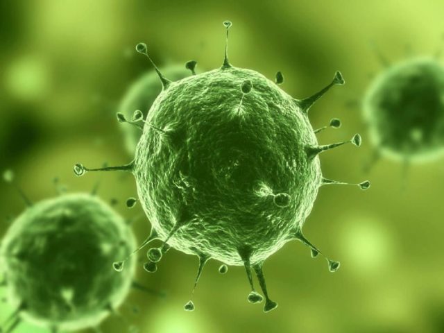
Something like this under an electron microscope the pathogens of cattle mycoplasmosis look
Reasons for infection
Calves are most sensitive to mycoplasmas, although cattle can be infected at any age. The main carriers of mycoplasmosis are sick and recovered cattle.
From sick animals, the pathogen is released into the external environment along with physiological fluids:
- urine;
- milk;
- discharge from the nose and eyes;
- saliva, including when coughing;
- other secrets.
Mycoplasmas get onto bedding, feed, water, walls, equipment, infecting the entire environment and being transmitted to healthy animals.
Also, infection with mycoplasmosis of cattle occurs in the "classical" ways:
- orally;
- airborne;
- contact;
- intrauterine;
- sexual.
Mycoplasmosis does not have a pronounced seasonality, but the greatest number of infections occurs in the autumn-winter period, when cattle are transferred to farms.
The area of distribution and the intensity of infection largely depend on the conditions of detention and feeding and the microclimate of the premises. Cattle mycoplasmosis stays in one place for a long time. This is due to the long period of preservation of bacteria in the body of recovered animals.
Symptoms of mycoplasmosis in cows
The incubation period lasts 7-26 days. Most often, symptoms of mycoplasmosis are observed in calves weighing 130-270 kg, but clinical signs may appear in adult animals. A clear manifestation of mycoplasmosis occurs only 3-4 weeks after infection. The disease spreads most rapidly in cold, wet weather and when cattle are overcrowded. The initial symptoms of mycoplasmosis are very similar to pneumonia:
- shortness of breath: cattle make every effort to draw air into the lungs and then push it out;
- frequent sharp cough, which can become chronic;
- discharge from the nose;
- sometimes conjunctivitis;
- loss of appetite;
- gradual exhaustion;
- temperature 40 ° C, especially if a secondary infection is "hooked" on mycoplasmosis;
- with the transition of the disease to the chronic stage, the temperature is only slightly higher than normal.
Arthritis begins a week after the onset of pneumonia. With arthritis in cattle, one or more joints swell. The mortality begins 3–6 weeks after the onset of clinical signs.
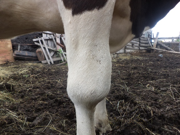
Arthritis in cattle is a "normal" phenomenon in mycoplasmosis
With the genital form of mycoplasmosis in cattle, abundant purulent discharge from the vagina is observed. The mucous membrane of the vulva is completely covered with small red nodules. A sick cow is no longer fertilized. Inflammation of the udder is also possible. In bulls, swelling of the epididymis and spermatic cord is determined by palpation.
Diagnosis of mycoplasmosis in cattle
Due to the similarity of the symptoms of mycoplasmosis with other diseases of cattle, the diagnosis can only be made by a comprehensive method. When determining the disease, take into account:
- Clinical signs;
- epizootological data;
- pathological changes;
- results of laboratory tests.
The main emphasis is placed on pathological changes and laboratory studies.
Pathological changes
Changes depend on the area of the main lesion by mycoplasmas. When infected by airborne droplets and by contact, the mucous membranes of the eyes, mouth and nasal cavity are primarily affected.
In case of eye disease, corneal opacity and its roughness are noted. The conjunctiva is edematous and reddened. As a result of an autopsy, most often in parallel with eye damage, hyperemia of the mucous membrane of the nasal passages is detected. Lesions in the middle and main lobes of the lungs are detected with a latent or initial course of the disease. The lesions are dense, gray or red-gray in color. The connective tissue is gray-white. In the bronchi, mucopurulent exudate. The bronchial walls are thickened, gray. Lymph nodes in the area of infection may be enlarged. When mycoplasmosis is complicated by a secondary infection, necrotic foci are found in the lungs.
The spleen is swollen. The kidneys are slightly enlarged, there may be hemorrhages in the renal tissue. Dystrophic changes in the liver and kidneys.
In the case of penetration of mycoplasmas into the udder, the consistency of its tissues is dense, the connective interlobular tissue is overgrown. Development of abscesses is possible.
When the genital organs are affected by mycoplasmosis, cows observe:
- swollen lining of the uterus;
- thickening of the fallopian tubes;
- serous or serous-purulent masses in the lumen of the oviducts;
- catarrhal-purulent salpingitis and endometritis.
Bulls develop epididymitis and vesiculitis.
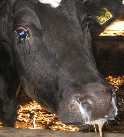
Discharge from the eyes and nose must be sent to the laboratory for analysis
Laboratory research
For samples, the following is sent to the laboratory:
- swabs from the cow's vagina;
- semen;
- embryonic membranes;
- milk;
- pieces of lungs, liver and spleen;
- bronchial lymph nodes;
- pieces of the brain;
- aborted or stillborn fetuses;
- the affected joints in general condition;
- flushes and mucus from the nose, provided that the upper respiratory tract is affected.
Tissue samples are delivered to the laboratory frozen or chilled.
For intravital diagnostics, 2 blood serum samples are sent to the laboratory: 1st when clinical signs appear, 2nd after 14-20 days.
Treatment of mycoplasmosis in cattle
Most antibiotics kill bacteria by attacking the cell wall.The latter is absent in mycoplasmas, so there is no specific treatment. For the treatment of mycoplasmosis in cattle, a complex system is used:
- antibiotics;
- vitamins;
- immunostimulants;
- expectorant drugs.
The use of antibiotics in cattle mycoplasmosis is due to the desire to prevent the complication of the disease by a secondary infection. Therefore, either drugs with a broad spectrum of action are used, or narrowly targeted: acting on microorganisms only in the gastrointestinal tract, lungs or genitals.
In the treatment of mycoplasmosis in cattle, the following are used:
- chloramphenicol (the main area of influence is the gastrointestinal tract);
- enroflon (broad-spectrum veterinary drug);
- antibiotics of the tetracycline group (used in the treatment of the respiratory and genitourinary systems and eye diseases).
The dose and type of antibiotic is prescribed by a veterinarian, since there are other drugs for mycoplasmosis that are not intended for the treatment of herbivorous cattle. The method of administration of a particular substance is also indicated by the veterinarian, but short instructions are usually also on the package.
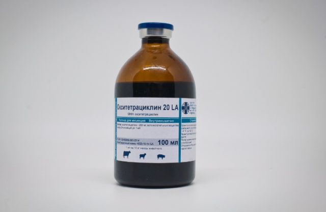
One of the antibiotics of the tetracycline group that can be used in the treatment of cattle mycoplasmosis
Prevention measures
Prevention of mycoplasmosis begins with standard veterinary rules:
- not to move animals from farms with mycoplasmosis;
- inseminate cows with only healthy sperm;
- do not introduce new individuals into the cattle herd without a month's quarantine;
- regularly carry out pest control, disinfection and deratization of premises where livestock are kept;
- regularly disinfect equipment and implements on the farm;
- provide cattle with optimal housing conditions and diet.
If mycoplasmosis is detected, milk from sick cows is subjected to heat treatment. Only then is it usable. Sick animals are immediately isolated and treated. The rest of the herd is monitored. Premises and equipment are disinfected with solutions of formalin, iodoform or chlorine.
Vaccinations are not carried out due to the lack of a vaccine against mycoplasmosis for cattle. So far, such a drug has been developed only for poultry.
Conclusion
Cattle mycoplasmosis is a disease that requires constant monitoring by the animal owner. The very case when it is better to once again mistake a simple clogged eyes for mycoplasmosis than to start the disease. The higher the concentration of the pathogen in the body, the harder it will be to cure the animal.


